Paper Art - Climbing x Synchrotron Microtomography
This is the first part in my series where I make a piece of art based on my most recent publication. This is also the image you see in the banner of this site! I made this piece after writing my first paper, the review paper about synchrotron microtomography in bone because at the time I was very interested in climbing. It’s very simple and a bit amateurish, but I have a strong attachment to this picture because it combines two of the things I discovered and learned about after coming to Utah from North Carolina.
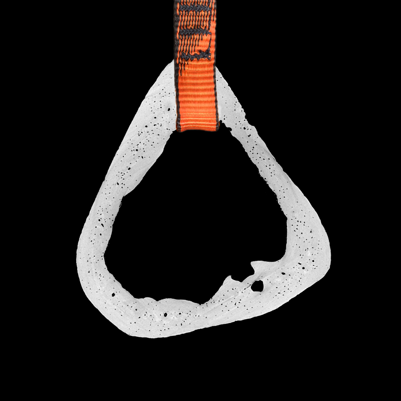
In light of the subject of the paper, I made the subject of this work a synchrotron microtomography scan image. More specifically, it’s a cross-section of a mouse femur. This scan is part of a dataset that I performed microstructure analysis on, which has yet to be published. All of the very small voids in the bone are the osteocyte lacunae. Osteocytes compose of 90% of cells in bone, and here you can visually see how numerous they truly are. Some of the slightly larger voids are canals that run through the bone to transport nutrients. It is actually quite difficult to discern canals from osteocyte lacunae in some cases if only a single cross-section is shown, such as here. Both the lacunae and the canals make up the greater lacunocanalicular network, which is responsible for local remodeling. Sometimes I get a bit tired of looking at the scans over and over, especially if the project is hard or if I run into unforeseen challenges. However, when I step back and look at even just one image like here and take in the detail, I remember how awesome it is for me to be able to work on this type of data.
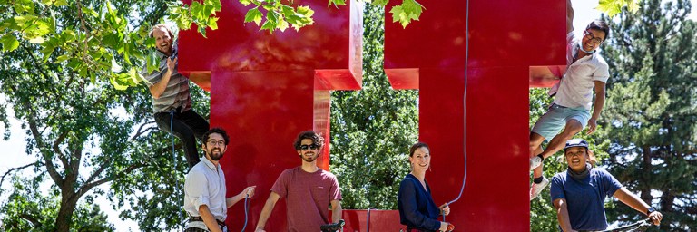
The other aspect of the image is the part of the quickdraw that would connect to the carabiner for clipping into a bolt in sport climbing. Part of the reason that I chose the bone cross-section that I did was that it reminded me of a carabiner in the first place. At first I wanted to add in the part that you would open to clip in, but in the end I did not want to hide any part of the bone to keep it as minimal as possible. I mentioned in the short description section that I am interested in climbing, but almost the entire lab is interested in climbing. I just got into climbing and bouldering, but I am really a novice. Mike, another Ph.D. student who works with micro-CT data as well is more experienced and has taken me climbing and bouldering a few times. Dr. Claire Acevedo, the head of the lab, is also very into climbing. Sometimes, we take lab climbing trips all together. Climbing has been an activity that really helped bring the lab together and make it feel like a close group.